

Simple columnar epithelium, 3D illustration. Histology background. Columnar epithelium is found in digestive system, uterus
Коллекция по умолчанию
Коллекция по умолчанию
Создать новую

Section of a dog ciliated columnar epithelium under the microscope.
Коллекция по умолчанию
Коллекция по умолчанию
Создать новую

Cross-section esophagus of the dog. Medical teaching equipment and biological tissue slide. Educational material for the study and treatment of doggy. Colouring with hematoxylin and eosin.
Коллекция по умолчанию
Коллекция по умолчанию
Создать новую

Pseudostratified columnar epithelium, 3D illustration. Epithelium found in trachea and upper part of digestive tract
Коллекция по умолчанию
Коллекция по умолчанию
Создать новую

Old color grunge vintage weathered background abstract antique texture with retro pattern
Коллекция по умолчанию
Коллекция по умолчанию
Создать новую

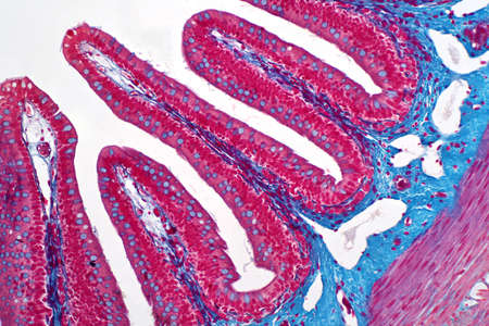
Small intestine with villi under the microscope 200x
Коллекция по умолчанию
Коллекция по умолчанию
Создать новую

A cross section of ciliated epithelium under the microscope.
Коллекция по умолчанию
Коллекция по умолчанию
Создать новую

Artery blood vessel under microscope view for education histology. Histological for human physiology.
Коллекция по умолчанию
Коллекция по умолчанию
Создать новую

Closeup view from above on heads of cotton buds laid in a horizontal line on pink background.
Коллекция по умолчанию
Коллекция по умолчанию
Создать новую

A close up of a very large piece of fruit with holes in it, AI
Коллекция по умолчанию
Коллекция по умолчанию
Создать новую

Old color grunge vintage weathered background abstract antique texture with retro pattern. Modern futuristic painted wall for backdrop or wallpaper with copy space. Close up image
Коллекция по умолчанию
Коллекция по умолчанию
Создать новую

Epithelium
Коллекция по умолчанию
Коллекция по умолчанию
Создать новую

Probiotics Lactobacillus acidophilus. Human microbiome background. 3d-rendering
Коллекция по умолчанию
Коллекция по умолчанию
Создать новую

Cross section of a cotton leaf under the microscope.
Коллекция по умолчанию
Коллекция по умолчанию
Создать новую

Showing Light micrograph of the Thyroid gland and Thymus gland human Child under the microscope for education in the laboratory.
Коллекция по умолчанию
Коллекция по умолчанию
Создать новую

Fibrinoid necrosis of the vessel wall, light micrograph, photo under microscope
Коллекция по умолчанию
Коллекция по умолчанию
Создать новую

Коллекция по умолчанию
Коллекция по умолчанию
Создать новую

Many porous red concrete surfaces
Коллекция по умолчанию
Коллекция по умолчанию
Создать новую

Human cerebellar cortex, photomicrograph showing Purkinje cells, granular cells, and molecular layer involved in motor coordination and balance.
Коллекция по умолчанию
Коллекция по умолчанию
Создать новую

Water lily with leaf in cross section 200x
Коллекция по умолчанию
Коллекция по умолчанию
Создать новую

Well-differentiated intestinal adenocarcinoma, light micrograph, photo under microscope
Коллекция по умолчанию
Коллекция по умолчанию
Создать новую

Saggy skin layer and skin cells, 3D rendering, beauty, and medicine
Коллекция по умолчанию
Коллекция по умолчанию
Создать новую

Drops- liquid foam. Fluid aqua- abstract pattern nature. Background- cleansing wash. Shampoo bubbles- soapy water
Коллекция по умолчанию
Коллекция по умолчанию
Создать новую

Anatomy and Histological Bone, Elastic cartilage human and Joint of human foetus under the microscope for education.
Коллекция по умолчанию
Коллекция по умолчанию
Создать новую

Ovarian cancer, light micrograph, photo under microscope. Photograph shows a fragment of a cancerous tumor in the female ovary. Selective focus
Коллекция по умолчанию
Коллекция по умолчанию
Создать новую

Root bacteria nodules in a bean root under the microscope.
Коллекция по умолчанию
Коллекция по умолчанию
Создать новую

diagonal lines of toned to pink cookies with ornament edge and symmetrical points on every of them. rows of few cookies lay one on one. cookies texture as sweet sugar background of homemade sweetness
Коллекция по умолчанию
Коллекция по умолчанию
Создать новую

Education anatomy and Histological sample of Human under the microscope.
Коллекция по умолчанию
Коллекция по умолчанию
Создать новую

Tissue of Small intestine (Duodenum) and Vermiform appendix Human under the microscope in Lab.
Коллекция по умолчанию
Коллекция по умолчанию
Создать новую

Tissue of Small intestine (Duodenum), Large intestine Human and Stomach Human under the microscope in Lab.
Коллекция по умолчанию
Коллекция по умолчанию
Создать новую

Renal tuberculosis, light micrograph, photo under microscope
Коллекция по умолчанию
Коллекция по умолчанию
Создать новую

Coral Porosity Detail: A detailed image highlighting the fine, porous patterns in the coral's texture.
Коллекция по умолчанию
Коллекция по умолчанию
Создать новую

Tissue of Small intestine (Duodenum), Large intestine Human and Stomach Human under the microscope in Lab.
Коллекция по умолчанию
Коллекция по умолчанию
Создать новую

Artery section under the microscope
Коллекция по умолчанию
Коллекция по умолчанию
Создать новую

Bacterial dysentery, light micrograph, photo under microscope showing accumulation of inflammatory cells, changes in structure of intestinal epithelium
Коллекция по умолчанию
Коллекция по умолчанию
Создать новую

Tissue of Small intestine (Duodenum) and Vermiform appendix Human under the microscope in Lab.
Коллекция по умолчанию
Коллекция по умолчанию
Создать новую

Bubble Tip Anemone - Entacmaea quadricolor.
Коллекция по умолчанию
Коллекция по умолчанию
Создать новую

Pork liver tissue cut 100x
Коллекция по умолчанию
Коллекция по умолчанию
Создать новую

Pandemic medical health, Virus background
Коллекция по умолчанию
Коллекция по умолчанию
Создать новую

Chronic pyelonephritis, light micrograph, photo under microscope
Коллекция по умолчанию
Коллекция по умолчанию
Создать новую

Prostate cancer, light micrograph, photo under microscope
Коллекция по умолчанию
Коллекция по умолчанию
Создать новую

Anatomy and Histological Bone, Elastic cartilage human and Joint of human foetus under the microscope for education.
Коллекция по умолчанию
Коллекция по умолчанию
Создать новую

Crocodile skin texture
Коллекция по умолчанию
Коллекция по умолчанию
Создать новую

3d rendered illustration of human cilia
Коллекция по умолчанию
Коллекция по умолчанию
Создать новую

Old color grunge vintage weathered background abstract antique texture with retro pattern. Modern futuristic painted wall for backdrop or wallpaper with copy space. Close up image
Коллекция по умолчанию
Коллекция по умолчанию
Создать новую

Intestine animal tissue under microscope view. histology of intestine.
Коллекция по умолчанию
Коллекция по умолчанию
Создать новую

Histopathology of human under microscope view for education in laboratory.
Коллекция по умолчанию
Коллекция по умолчанию
Создать новую

Pink wave of liquid vortex with bubbles
Коллекция по умолчанию
Коллекция по умолчанию
Создать новую

Education anatomy and Histological sample of Human under the microscope.
Коллекция по умолчанию
Коллекция по умолчанию
Создать новую

Abstract pink watercolor on paper texture can use as background
Коллекция по умолчанию
Коллекция по умолчанию
Создать новую

Cervical erosion, also known as eversion and ectropion, light micrograph. Photo under microscope, selective focus. Cervical ectopy
Коллекция по умолчанию
Коллекция по умолчанию
Создать новую

Microscopic image of the eggs parasite (Toxocara canis) under the microscope view for education
Коллекция по умолчанию
Коллекция по умолчанию
Создать новую

Abstract background. Pink wall, porous stone texture
Коллекция по умолчанию
Коллекция по умолчанию
Создать новую

Host cells with spores (mold) are inside wood under the microscope for education.
Коллекция по умолчанию
Коллекция по умолчанию
Создать новую
Vibrant abstract cellular texture
Коллекция по умолчанию
Коллекция по умолчанию
Создать новую

Uterine cancer, light micrograph, photo under microscope
Коллекция по умолчанию
Коллекция по умолчанию
Создать новую

Grungy pink cement wall texture. Abstract background and texture for design.
Коллекция по умолчанию
Коллекция по умолчанию
Создать новую

Bacteria, Bacterial colony, Microbes, Salmonella Bacteria
Коллекция по умолчанию
Коллекция по умолчанию
Создать новую

human sperms fine with microscope in laboratory.
Коллекция по умолчанию
Коллекция по умолчанию
Создать новую

Bowen's Disease Tumor under the microscope 100x
Коллекция по умолчанию
Коллекция по умолчанию
Создать новую

Artery blood vessel under microscope view for education histology. Histological for human physiology.
Коллекция по умолчанию
Коллекция по умолчанию
Создать новую

Image of multiple colourful brushes moving in formation and changing colours. abstract colour and movement concept digitally generated image.
Коллекция по умолчанию
Коллекция по умолчанию
Создать новую

Old color grunge vintage weathered background abstract antique texture with retro pattern. 16:9 aspect ratio
Коллекция по умолчанию
Коллекция по умолчанию
Создать новую

Human fat body tissue under microscope view for physiology education.
Коллекция по умолчанию
Коллекция по умолчанию
Создать новую

Histology of human kidney, light micrograph showing nephron. Microscopy, hematoxylin and eosin staining
Коллекция по умолчанию
Коллекция по умолчанию
Создать новую

Atypical squamous epithelial hyperplasia, photomicrograph showing thickened stratified epithelium with nuclear atypia and basal layer crowding.
Коллекция по умолчанию
Коллекция по умолчанию
Создать новую

Villi of small intestine, 3D illustration. Intestinal environment, close-up view
Коллекция по умолчанию
Коллекция по умолчанию
Создать новую
close-up of pink brain coral pattern, created with generative ai
Коллекция по умолчанию
Коллекция по умолчанию
Создать новую

Abstract background. Germs microorganism cells under microscope. Fast multiplication of bacteria. Infection and microbe. Red coloured. Microbiology, popular scientific background. 3D Rendering
Коллекция по умолчанию
Коллекция по умолчанию
Создать новую

Close up of pink carpet texture
Коллекция по умолчанию
Коллекция по умолчанию
Создать новую

Vials for veterinary use to cure different diseases. No people
Коллекция по умолчанию
Коллекция по умолчанию
Создать новую

Anatomy and Histological Bone, Elastic cartilage human and Joint of human foetus under the microscope for education.
Коллекция по умолчанию
Коллекция по умолчанию
Создать новую
Colon biopsy from the colonoscopy. The pathology report describes normal colonic mucosa fragment with colic glands.
Коллекция по умолчанию
Коллекция по умолчанию
Создать новую

Histology of human stomach, fundic region. Light micrograph, isolated on white background, hematoxylin and eosin staining
Коллекция по умолчанию
Коллекция по умолчанию
Создать новую

Abstract geometric pattern with blocks. Randomly ordered angle. Movable structure.
Коллекция по умолчанию
Коллекция по умолчанию
Создать новую

Benign prostatic hyperplasia. Micrograph shows dilated glands, papillary projections inside the lumen of the glands, cystic dilatation with accumulation of secretory material. Photo under microscope
Коллекция по умолчанию
Коллекция по умолчанию
Создать новую

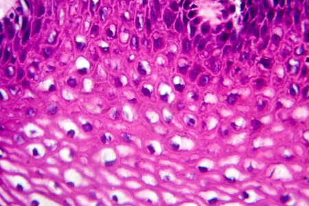
Human tongue section showing papillae and stratified epithelium
Коллекция по умолчанию
Коллекция по умолчанию
Создать новую

science botany micrograph plant root tip tissue cell, Magnification 200X
Коллекция по умолчанию
Коллекция по умолчанию
Создать новую

Macrophotography of fabric texture abstract background close up view.
Коллекция по умолчанию
Коллекция по умолчанию
Создать новую

Purple water drink bubbles abstract background
Коллекция по умолчанию
Коллекция по умолчанию
Создать новую
Vibrant abstract underwater tunnel
Коллекция по умолчанию
Коллекция по умолчанию
Создать новую

Human liver tissue under microscope view
Коллекция по умолчанию
Коллекция по умолчанию
Создать новую

Renal cell carcinoma, light micrograph, photo under microscope
Коллекция по умолчанию
Коллекция по умолчанию
Создать новую

Fruits of raspberry background.
Коллекция по умолчанию
Коллекция по умолчанию
Создать новую

Close up image of a strawberry as background
Коллекция по умолчанию
Коллекция по умолчанию
Создать новую

Mechanical Science as a Art Background Concept
Коллекция по умолчанию
Коллекция по умолчанию
Создать новую

Plant stem with sieve cells under the microscope.
Коллекция по умолчанию
Коллекция по умолчанию
Создать новую

Acute myocardial infarction, histology of heart tissue, light micrograph. Area of infarct is paler than than the relatively viable area of heart muscle
Коллекция по умолчанию
Коллекция по умолчанию
Создать новую

Anatomy and Histological Epididymis and Testis human cells under microscope.
Коллекция по умолчанию
Коллекция по умолчанию
Создать новую
Atypical squamous epithelial hyperplasia, photomicrograph showing thickened stratified epithelium with nuclear atypia and basal layer crowding.
Коллекция по умолчанию
Коллекция по умолчанию
Создать новую

Lichen, Fungus, cross section slide under the microscope view for education biology.
Коллекция по умолчанию
Коллекция по умолчанию
Создать новую

Cell membrane structure background, 3d rendering. Digital drawing.
Коллекция по умолчанию
Коллекция по умолчанию
Создать новую

Squamous cell carcinoma, light micrograph, photo under microscope
Коллекция по умолчанию
Коллекция по умолчанию
Создать новую

the abstract colors and blurred background
Коллекция по умолчанию
Коллекция по умолчанию
Создать новую
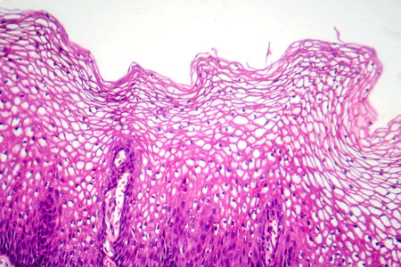
Atypical squamous epithelial hyperplasia, photomicrograph showing thickened stratified epithelium with nuclear atypia and basal layer crowding.
Коллекция по умолчанию
Коллекция по умолчанию
Создать новую

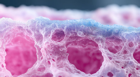
Histopathology of lung emphysema, light micrograph, photo under microscope showing enlargement of air spaces in lung tissue and destruction of alveolar septa
Коллекция по умолчанию
Коллекция по умолчанию
Создать новую

Liver cyst formation, light micrograph, photo under microscope
Коллекция по умолчанию
Коллекция по умолчанию
Создать новую

Section of the pyloric region of a dog stomach under the microscope.
Коллекция по умолчанию
Коллекция по умолчанию
Создать новую

Histopathology of pneumonia, light micrograph, photo under microscope. Cellulose aspiration pneumonia
Коллекция по умолчанию
Коллекция по умолчанию
Создать новую

Education anatomy and Histological sample Striated (Skeletal) muscle of mammal Tissue under the microscope.
Коллекция по умолчанию
Коллекция по умолчанию
Создать новую
Legion-Media
Создайте свои проекты на основе качественных стоковых фотографий и видео.


Copyright © Legion-Media.